


Osteochondrosis is a chronic disease symptomatically expressed by dystrophic disorders in articulated cartilage. Osteochondrose of spine usually occurs, when the changes occur in interviolent discs and interface joints. Depending on the localization, thunder, thoracic and lumbar osteochondrosis are different. Osteochondrosis is also often located, in which all parts of the spine suffer. Pathology requires consulting with a doctor and an integrated approach to treatment.
Description
Spine diseases, as well as other chronic diseases, are quickly "become younger. "If the earlier pain in the back and joints bothered the elderly, today the patients from 18-30 years are increasingly treated by doctors.
Scientists believe that the directness of a person is a prerequisite for the development of this disease, osteochondrose is also facilitated by an extended sitting apartment. In addition, with age, articular cartilage loses elasticity and elasticity, thin out; Intervertebral Disks lose moisture and the ability to absorb the impact, become vulnerable at the time of physical effort.
Reasons
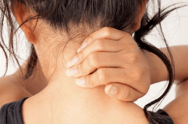
The causes of cervical osteochondrose lie in loads and they affect the cervical region under different circumstances. Therefore, the muscles in the neck starts intensively reducing, thus compensating for that way, as a result of spasm, as well as blood flow violation in this area.
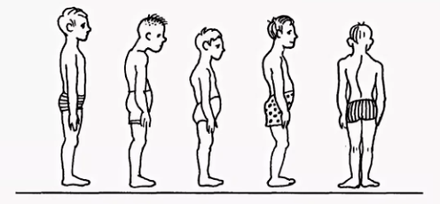
- Breach of injury for scoliolose, stop, round back, kyphosis and other disorders, even if lies, cause serious breach of the spinal stair. As a result, the load on intervebrebral discs was distributed unevenly, which causes their deformation and increased wear. Ruminums are beginning to approach, causing violation of nerve processes, cervical osteochondrosis develops quite fast. Similar consequences have a violation of the position caused by changing the natural position of the ribs.
- Savings muscle spasmodic reaction muscles back, breast, the press can lead to the fact that individual body parts are very tense. As a result, the general equilibrium body position is disturbed, causing the change in the position of the spine. Deformations may affect the region of Grlić's registry or other parts of the spinal column, causing osteochondrose of the chest, cervical and lumbar parts.
- Violation of blood supply, because vertebrates do not have a direct connection with the circulatory system, they receive nutrition from the environment. Violation of blood supply to cervix leads to the fact that discs do not receive sufficient reaction fluids (renovation of the form due to moisture absorption) and crunchy renovation. As a result, their wear is accelerated, the decline in the distance between the vertebral region is accelerated, leading to osteochondrose.
- Breach of innovation, reducing the sensitivity of nerve roots leads to pathological changes in their structure, as a result that the displacement and deformation of the cervical region remain unnoticed by the patient. After all, the pain is absent because of the sensitivity disorder.
- Internal bodies diseases are the wrong position of internal bodies, their displacement and descending due to different dysfunction lead to a violation of the general balance in the body. As a result, this sharply affects the position of the spinal column - cervix, lumbar vertebrates are displaced and deformed, leading to appropriate osteochondrose species.
In general, osteochondrosis of the cervical region develops due to the effects of negative external factors that violate the position of the natural balance of the spinal column and other systems of the human body.
Diagnostics
The diagnosis of cervical osteochondrosis begins by collecting all the necessary information about the patient. The specialist asks the complaints that bothering persons, interest in his professional activities, as well as how he spends his weekend. The important thing is the presence of osteochondrose in parents, grandparents, because this is a disease of hereditary nature.
Then the doctor continues directly to a visual examination of the patient. The cervical compartment and back is studied in the curvature of the position, palpatury cervical regions. This allows the expert to assess the degree of disease development, because in advanced cases, the palpation of the cervical region causes harsh pain.
You should pay attention to:
- on the severity of the cervix;
- shoulder height in the patient;
- Possibility of the asymmetry of the Supraclock area;
- Possibility of the door asymmetion (for example, the consequence of innate pathology or sharp muscle spasm);
- The state of the shoulder and upper limb muscles (for example, one-a-year muscle atrophy may indicate the compression of the spine of garden spine);
- The location of the beard - the beard is normal should be located along the middle line;
- Door movement (bending extinguishing, tilting rights and rotation and rotation).
Palpation is done in the initial position of the patient:
- lying on back;
- lying on the stomach;
- Sitting in a chair.
A study on the amount of movement is also implemented. It is performed in the initial position of a patient sitting on a chair (in order to improve another spine).
Distinguish the following basic movements in the cervical region:
- flexion;
- expansion;
- slopes on the right and left;
- Rotation.
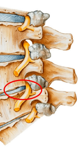
Approximately half of the flexion and the volume of the extension occurs between the back of the head, the vernival C1 and C2. The rest of the movement is done due to the basic vertebrae, with a large range of movements in the C5-C7 vertebral. The side slopes are evenly distributed between all vertebrae.
In order to precisely make a diagnosis, additional studies are prescribed:
- X -ray cervical regions. This method is suitable in the early stages of the disease, but can be useless in advanced forms.
- CT (computer tomography). It allows you to see structural changes in vertebrates, but with the help of this method, it is impossible to determine the size of the rain between vertebral.
- MRI. It is considered a most effective method of diagnosing cervical osteochondrose. You can determine the size of the hernia between the disks, as well as the degree of their development.
- The doctor can also prescribe a duplex scan that allows you to determine the violation of normal blood circulation in the arteries.
Treatment
Treatment of cervical osteochondrone is a complex therapy that involves taking drugs, gels, as well as different physiotherapy measures. Important role Game Therapeutic gymnastics, as well as massage of the problematic area.
Cure
It is impossible to eliminate the consequences of degenerative dystrophic changes in the spine without using drugs. The use of drugs in the treatment of osteochondrose of the Cervical region is one of the main points of a complex approach to healing, which use most traditional medicine specialists.
It is designed to solve several problems including:
- stop the symptom of pain and eliminate the inflammatory process;
- remove muscle cramp;
- encourage the process of regeneration of cartilage cells and bone tissue;
- strengthen body protective properties, increase immunity;
- Improve the general state by eliminating other symptoms that interfere with recovery.
Depending on these purposes, all drugs prescribed by a doctor suffering from cervical osteochondorosis can be conditionally divided into the following groups:
- Analgesics (nonsteroid medications that relieve pain).
- Anti-Aparnatory (steroid) are hormonal drugs that relieve inflammatory phenomena and which removes pain.
- Chondroprotectors are drugs that contain substances that replace cartilage components - hondroitine, hyaluronic acid.
- Musorelaxans. These are drugs that relax the muscle tone. They are used in surgery and orthopedics as ancillary medications to stop pain. Such drugs are managed by Parelet, and thus always under the supervision of the doctor.
- Vitamins. With the osteochondrose of the cervical region, the vitamins are prescribed, in the use of the peripheral nervous system and the improvement of conductivity. Votamini vitamins: B1, B6, B12, Fat-beans Vitamins: A, C, D, E. In recent years, medications containing both hospitals and vitamin components more often.
- Fats and gels for outdoor use.

Physiotherapy
The main goal of physiotherapy treatment is to encourage the process of regeneration in the body and eliminate pain. The most popular methods in the treatment of cervical chondrosis are as follows:
- Ultrasound. Phyimuim with ultrasonic waves is used to mitigate serious pain manifestations and inflammatory reactions. The ultrasound is a massage of the tissue door, after which the metabolism is activated.
- Vibration massage. Impact on the surface of the pain during the vibration massage is through mechanical oscillatory movements. A vibro tape bar is usually used for proper behavior of this physiotherapy.
- Electrophoresis. This method of conducting physiotherapy care for cervical osteochondrosis is done using diadgia and modulated currents, as well as electric fields when the body applies in medicine tissues. Electrophoresis perfectly relieves spasmodic syndrome and eliminates pain in inflamed muscles.
- Magnetotherapy. The essence of magnetotherapy as physiotism in the osteochondrose of the cervical region is explained by the use of constant or fields with magnet, frequency of different sizes. This method can help the patient remove pain and stop the inflammatory process on the hearth. The procedure is often done at home after gaining a special device for a magnetograph.
- Dutoenzor-therapy. Currently a pretty popular physiotherapy method, which consists of stretching the spinal column under the mass of the patient's body. In order to perform such a procedure, a mattress is required in a special way, which has sloping ribs and they change the location under the weight of your own body. The muscle tone is normalized, leading to their relaxation.
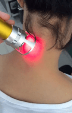
- Laser therapy. The laser has a complex effect on focus inflammation, activates biological processes in the tissues of the nervous system. This allows you to achieve a positive effect from treatment. The complex effect on the body consists in anti-intentional, analgesic and wound effect. One processing process of laser treatments must not exceed 15 minutes. This is the optimal time of Sector Exposure to Helić-Neon laser in the affected areas. In this case, the duration of lasers on one pain must not be more than 2 minutes.
- Balneotherapy. The benefits of mineral water have been known for a long time, that is what balneotherapy is based. The procedure implies the active use of water resources in the treatment of osteochondrose. In addition to the adoption of the bathroom, different types of souls and active swimming in the pool, therapy includes the use of the application of therapeutic muds with painful areas of the body. The therapeutic effect is achieved by the simultaneous effect of chemical active substances contained in water in different temperature conditions. The technique allows you to stop syndrome pain by improving local microcirculation in the tissues.
Practice therapy
It should be borne in mind that exercise therapy is not implemented when the signs of deterioration begin: pain. After the LFK complex, I can enhance and cause inconvenience.
There are several general billing recommendations:
- Physical education should take place indoors with good ventilation, excellent option on the street.
- Classes are carried out only in the period of remission of the disease (when there is no symptoms).
- Clothing in exercise therapy should be wide, not embarrassing movements and breathing.
- All movements are smooth, amplitude and repetition number gradually increases.
- If pain starts, you should immediately stop the lesson.
- Precedes classes and finish pressure and pulse measurements. When these indicators differ from the normal, the cargo should be reduced.
- It is advisable to listen to your breathing during the lesson, it will increase efficiency. All stretching exercises are performed on exhalation.
- It is very important to gradually increase the load and number of repetitions, it will reduce the risk of injury and prevent excessive work.
- Exercises are important to perform regularly, so you can achieve a quick result.
- Before starting an independent class, you must contact your doctor and agree with it a set of exercises.
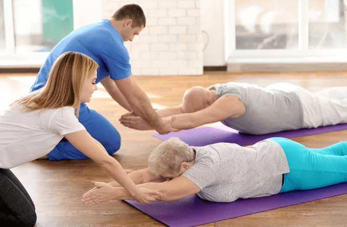
Recommended exercises in the initial position lie on the stomach:
- The head is eventually headed, hands on the back of the head, elbows in parallel from the floor. Raise your head with your hands from the floor, hold this position on 4 accounts, lower and relax. Repeat 2-4 times.
- The head is on the station on the chin, palms under his chin. Time time, stretch your hands ahead, two - expand yourself to the sides, three - stretch forward, four - starting position. Repeat 2-4 times.
- Hands extended forward. Swimming style "Rabbit", repeatedly 4-8 times.
- The palms under his chin, emphasis on the palm of your forehead. Alternately, take out the fifth buttocks. Repeat 4-8 times.
Recommended exercises in the initial position lie on the side (on the right, then on the left):
- Right hand is expanding, right ear is on it, raise your right hand with your head, hold the position on 4 accounts, the lower and relax. Repeat 2-4 times.
- The left hand lenses on the floor in front of the chest, left leg makes fly movements back and forth. Repeat 6-8 times.
- Left hand with the body, raise your left hand up and out, lower, lower. Repeat 2-4 times.
- Left handed to thigh. Drag both knees to your chest on exhale, correct your legs on inspiration. Repeat exercises 2-4 times.